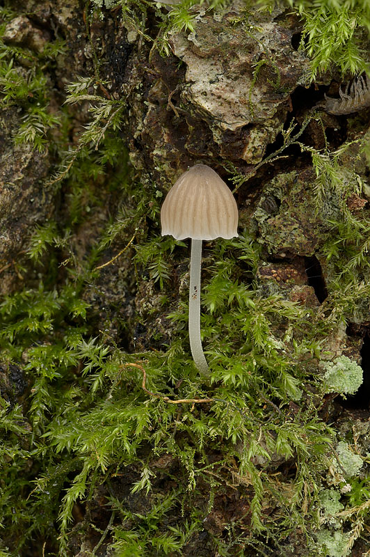
23 December 2025 Crab Wood, Hampshire. Photograph copyright Leif Goodwin. Cap Convex, usually with a small bump at the centre, pale brown, radially lined, paler towards the margin, margin scalloped, to about 10 mm across Gills Adnate to decurrent, whitish Stem Equal, smooth, translucent, whitish, to about 3 cm high Flesh White Smell Indistinct Taste Indistinct Season Autumn to winter Distribution Rarely reported Habitat Among moss on the bark of living deciduous trees Spore Print White Microscopic Features Spores ellipsoidal, smooth (6-10) x (5-7) µm2. Gill edge cystidia spindle to bladder shaped. Stem cystidia Edibility Inedible Notes The 2025 collection was independently identified by Eric Janke and Leif Goodwin. Both noted the absence of gill edge cystidia. A sample will be DNA sequenced. 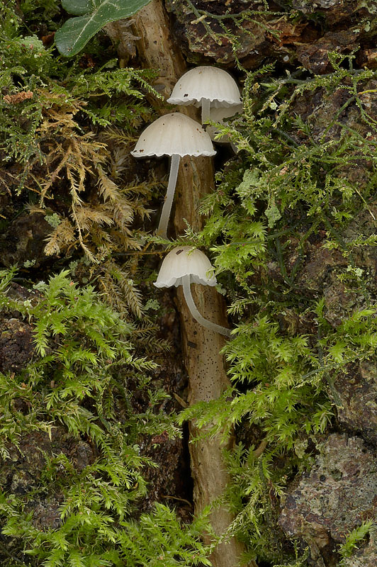
23 December 2025 Crab Wood, Hampshire. Photograph copyright Leif Goodwin. 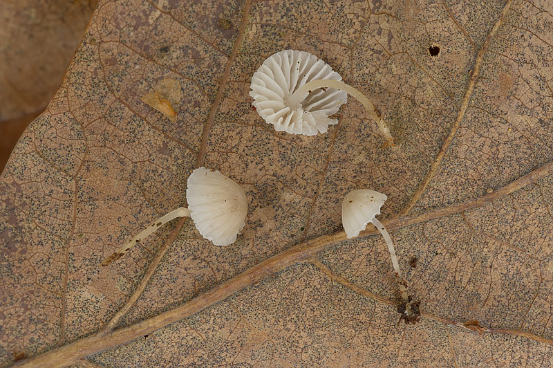
23 December 2025 Crab Wood, Hampshire. Photograph copyright Leif Goodwin. 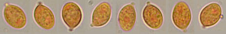
Spores in Congo Red solution viewed with a 100X immersion objective. 23 December 2025 Crab Wood, Hampshire. Photograph copyright Leif Goodwin. 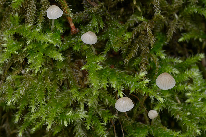
26 October 2013 Hampshire. Photograph copyright . 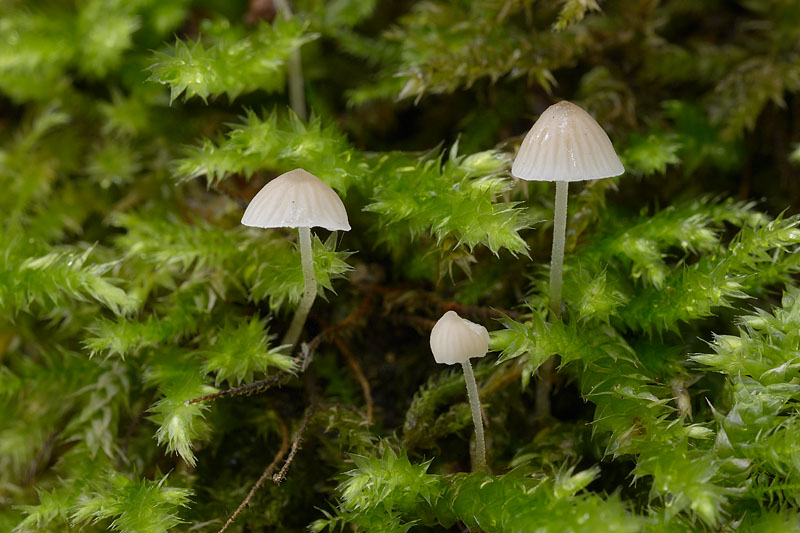
26 October 2013 Hampshire. Photograph copyright Leif Goodwin. 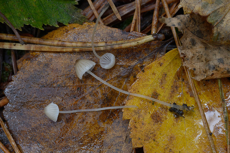
26 October 2013 Hampshire. Photograph copyright Leif Goodwin. 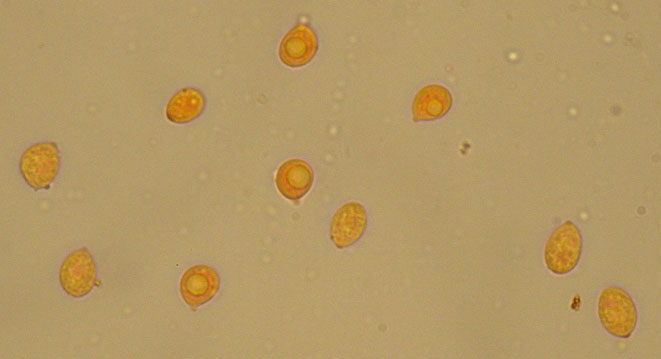
Spores in Congo Red solution viewed with a 100X immersion microscope objective. 26 October 2013 Hampshire. Photograph copyright Leif Goodwin. 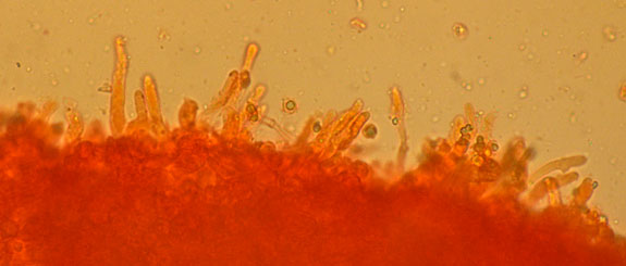
Gill edge cystidia viewed with a x40 microscope objective. 26 October 2013 Hampshire. Photograph copyright Leif Goodwin. |